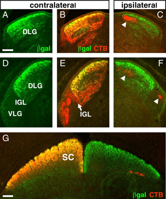
Figure 3.

Innervation of the adult lateral geniculate and superior colliculus by Brn3a RGCs. Alexa-594 CTB was injected unilaterally, and animals were killed after 3 d. Expression of the tauLacZ transgene was visualized with immunofluorescence for β-galactosidase. Brn3a RGC axons predominantly innervate the superficial layer of the DLG and are absent from the principal part of the VLG and all ipsilateral LG fibers. A–C, Retinal projections to the rostral lateral geniculate. D–F, Innervation of the caudal lateral geniculate. G, Retinal fibers innervating the superior colliculus. The arrowheads in C and F indicate ipsilateral fibers, which are devoid of β-galactosidase immunoreactivity. βgal, β-Galactosidase; SC, superior colliculus. Scale bars: (in A, G) 100 μm.
