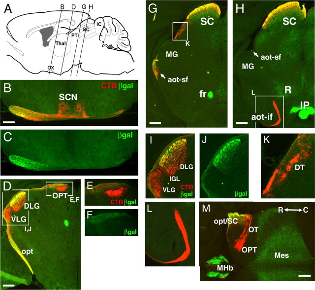
Figure 4.
Restricted targets of Brn3a RGCs in the thalamus and midbrain. A Brn3atLacZ mouse was enucleated unilaterally at P2, and the remaining eye was injected with CTB 2 d before the mice were killed at P30. Except for B and C, only the contralateral side is shown. A, Planes of coronal section for views A–L. B, C, Section immediately posterior to optic chiasm shows that retinal projection to the SCN is devoid of β-galactosidase immunoreactivity. D–F, I, J, Section at level of the caudal lateral geniculate and pretectum. The pretectal nuclei and the principal part of the VLG show no β-galactosidase immunoreactivity. G, K, Section through the rostral superior colliculus. H, L, Section near the middle of the superior colliculus, showing the accessory optic tract, which does not exhibit β-galactosidase immunoreactivity. M, Horizontal section through the midbrain and pretectal area. Aot, Accessory optic tract (sf, superior fasciculus; if, inferior fasciculus); DT, dorsal terminal nucleus of the accessory optic tract; fr, fasciculus retroflexus; IC, inferior colliculus; IP, interpeduncular nucleus; Mes, mesencephalon (deep layers of SC and central gray); MHb, medial habenula; MG, medial geniculate; opt, optic tract; OPT, olivary pretectal nucleus; OT, nucleus of the optic tract; ox, optic chiasm; PT, pretectum; R, red nucleus; SC, superior colliculus; Thal, thalamus; βgal, β-galactosidase; R, rostral; C, caudal. Where possible, nomenclature is adopted from Paxinos and Franklin (2001). Scale bars: B, 200 μm; D, G, H, M, 400 μm.

