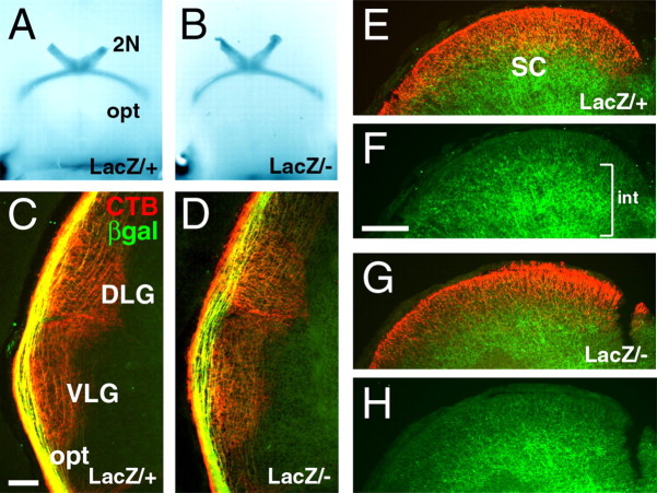
Figure 6.
Development of the retinothalamic/retinocollicular tracts in Brn3a null mice. A, B, Xgal staining of the optic chiasm at E16.5 in whole-brain preparations. C–G, Viable E18.5 embryos were injected with CTB unilaterally and killed after 8 h. C, D, CTB-labeled retinal axons can be seen in the lateral geniculate, but β-galactosidase-immunoreactive fibers remain mostly in or parallel to the optic tract. E–H, In the E18.5 midbrain, extensive labeling of intrinsic (int) midbrain Brn3a neurons is observed in deep layers of the superior colliculus(F,bracket), but few axons of the Brn3a RGCs have reached the superior colliculus at this stage in either Brn3atLacZ/+ (E, F) or Brn3atLacZ/– (G, H) embryos. 2N, Optic nerve; opt, optic tract; SC, superior colliculus; βgal, β-galactosidase. Scale bars: (in C) C–D, 100 μm; (in F) E–H, 200 μm.

