
Figure 8.
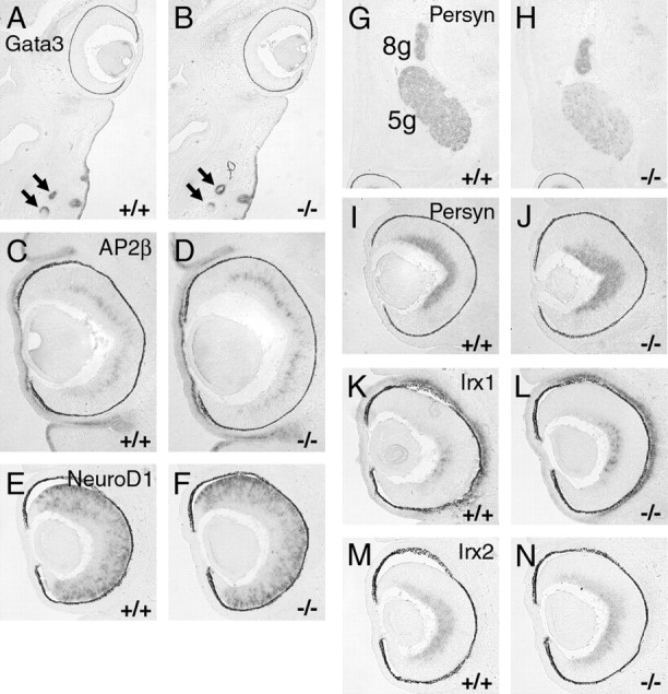
Expression of known Brn3a and Brn3b regulatory targets in the Brn3a–/– retina examined by in situ hybridization. Horizontal sections of E14.5 embryos are shown in all views, and the caudal/occipital part of the head or temporal side of the retina is at the top of each view. A, B, GATA3 is expressed strongly in whisker follicles (arrows) but not in the control or knock-out retina. C–F, Targets of Brn3a regulation in the trigeminal ganglion AP2β and NeuroD1 are unaltered in the Brn3a knock-out retina. G–J, Brn3b regulatory target persyn is decreased in the trigeminal ganglion but not in the retina of Brn3a null mice. K–N, Retinal expression of Irx1 is increased in the absence of Brn3a, but Irx2 shows no significant change. 5g, Trigeminal ganglion; 8g, vestibulocochlear ganglion.