Figure 4.
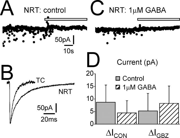
NRT neurons do not exhibit tonic GABAA current. A, Trace recorded from an NRT neuron under control conditions. Focal GBZ application (white bar) does not cause an outward shift in baseline current (ΔIGBZ = 3.4 pA; ΔICON = 8.9 pA). B, The waveform of the average IPSC for the same cell as in A. The average IPSC of a dLGN TC neuron has been scaled for comparison. Note that the IPSC of the NRT neuron has a characteristically longer decay than that of the TC neuron. C, Trace recorded from a different NRT neuron in the presence of extracellular GABA (1 μm). Focal application of GBZ still does not cause an outward shift in baseline current (ΔIGBZ = 9.8 pA; ΔICON = 6.5 pA). D, Graph showing the comparison of ΔICON and ΔIGBZ for NRT neurons under control conditions (gray columns) and in the presence of 1 μm GABA (hatched columns). Under neither condition is a tonic current apparent (Student's paired t test). Calibration in A also applies to C. Calibration in B applies only to the trace from the NRT neuron.

