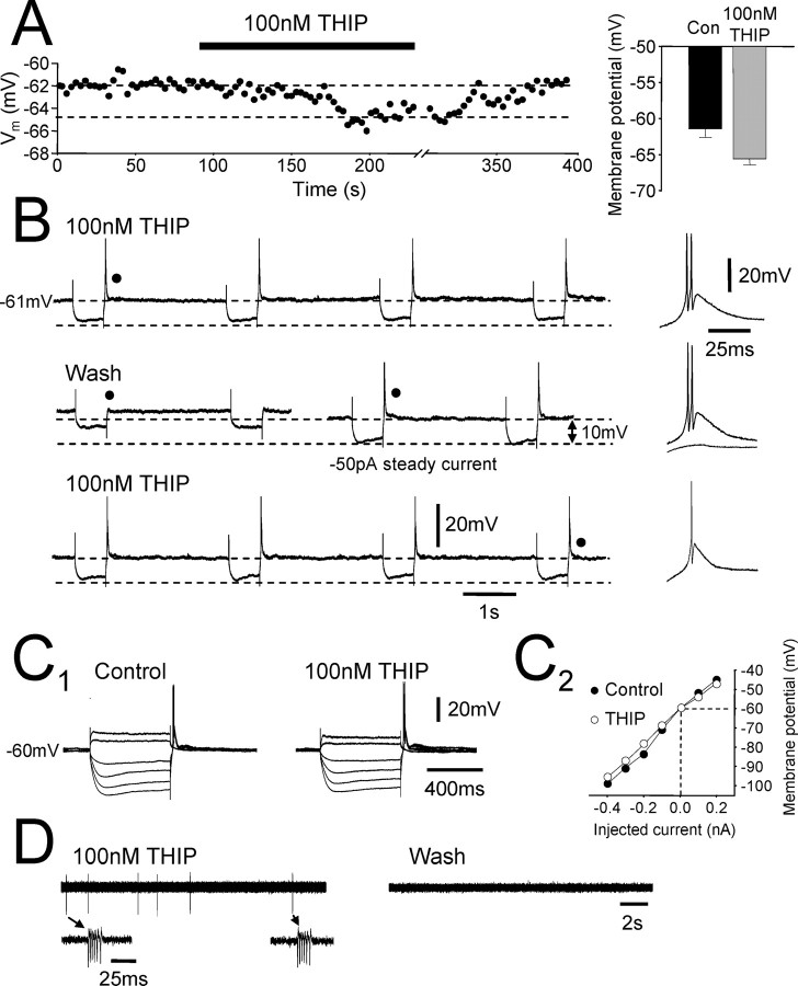
Figure 8.
Enhancement of the tonic current hyperpolarizes TC neurons and promotes burst firing. A, Intracellular current-clamp recording from a dLGN TC neuron. Bath application of 100 nm THIP (black bar) caused a small hyperpolarization (∼3 mV) that could be reversed during washout. The bar graph to the right shows the membrane potential before (black column) and after (gray column) 100 nm THIP application for all recorded neurons. B, Recording from another dLGN TC neuron showing membrane potential responses to hyperpolarizing current steps (–100 pA) during 100 nm THIP application (top trace), wash (middle traces), and reapplication of THIP (bottom trace). Note how removal of THIP from the aCSF results in a small depolarization and increase in input resistance that is reversed after reapplication of THIP. To the right are the membrane responses at the offset of the current steps, as indicated (•). C1, Response of a dLGN TC neuron to depolarizing (100 and 200 pA) and hyperpolarizing (–400, –300, –200, and –100 pA) current steps under control conditions (left) and after bath application of 100 nm THIP (right). C2, Current–voltage plot for the neuron in C1. Values were taken from the maximum deflection of the membrane potential during the current steps. THIP application causes a small decrease in apparent input resistance in response to hyperpolarizing current steps. D, Extracellular single-unit recording of a dLGNTC neuron in the presence of 100 nm THIP (left) and after wash (right). Only burst firing is apparent in the presence of THIP (see insets), whereas its removal from the aCSF results in the silencing of the unit.

