
Figure 3.
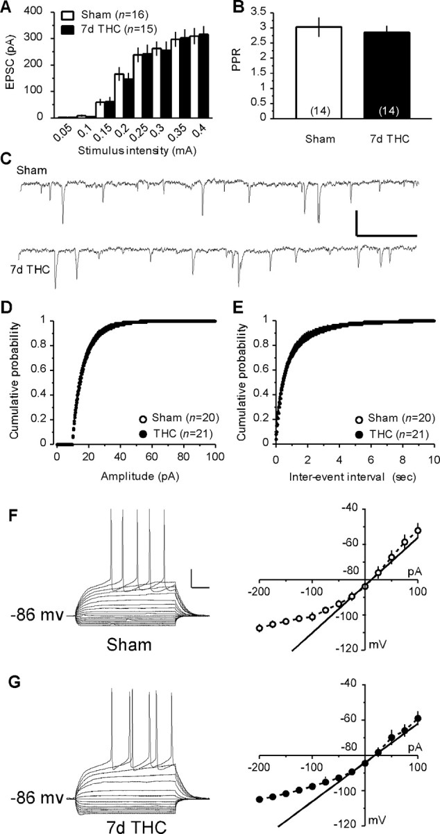
Repeated exposure to THC alters neither basal synaptic strength and release probability at the PFCx–NAc synapses nor the basic electrophysiological properties of MSNs. A, Similar input–output curves were obtained when stimulating PFCx afferences and recording the EPSCs evoked in accumbens MSNs from vehicle-treated (sham, n=16) and THC-treated (7d THC, n=15) mice.B, The PPR of the EPSCs did not differ between the MSNs from sham (n=14) and 7 d THC-treated (n = 14) mice. C, Representative 3 s sweeps showing the sEPSCs recorded from MSNs in the NAc of sham and 7 d THC-treated mice. Calibration: 20 pA (y-axis), 200 ms (x-axis). D, E, The distribution of the sEPSCs amplitude (D) and interevent interval (E) did not differ between MSNs from sham (n = 20) and 7 d THC-treated (n = 21) mice. F, G, Typical responses to hyperpolarizing and depolarizing somatic current pulses of an MSN in the NAc of a sham (F) and a 7 d THC-treated (G) mouse. Calibration: 20 pA (y-axis), 50 ms (x-axis). Similar average I–V curves were calculated from MSNs recorded in the NAc of sham (n = 8) and THC-treated (n = 11) mice.