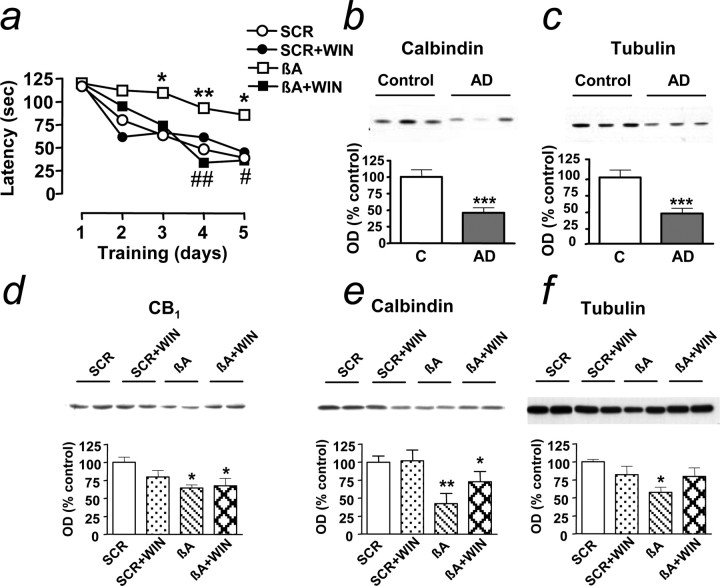
Figure 5.
Cannabinoid treatment prevents cognitive impairment and loss of neuronal markers in rats. a, Latency (in seconds) to find a hidden platform in the water maze during training. Results are mean of n = 5 in each group; SEM have been omitted for clarity and were always <12% of the mean; *p < 0.05 and **p < 0.01 compared with SCR-treated rats at the same training day; #p < 0.05 and ##p < 0.01 compared with βA-treated rats (ANOVA with Bonferroni's post hoc test). WIN, WIN55,212-2. b, c, Expression of calbindin (b) and α-tubulin (c) in control (C) and AD frontal cortex; results are mean ± SEM of n = 18 control and AD; ***p < 0.001 compared with controls. d-f, Expression of CB1 (d), calbindin (e), and α-tubulin (f) in frontal cortex of rats at 2 months after treatment. OD, Optical density. Results are mean ± SEM of n = 5 in each group; *p < 0.05 and **p < 0.01 compared with SCR-treated rats (ANOVA with Bonferroni's post hoc test); representative blots are shown. Error bars represent SEM.

