Figure 4.
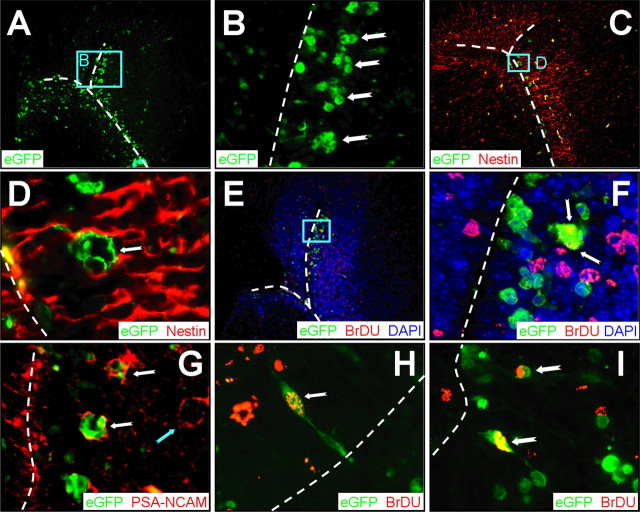
CVB3 infects type A, as well as type B, stem cells. Transverse paraffin-embedded sections were deparaffinized and immunostained using antibodies against nestin, BrDU, or PSA-NCAM. The ependymal cell layer is represented by the dashed white line. A, Many clusters of infected cells (eGFP+) were observed within the subventricular zone. B, Higher magnification (A, cyan box) revealed the dense clusters of infected cells (notched arrows) morphologically similar to type A neuronal progenitor cells within the SVZ. C, As expected, nestin staining (red) was observed near the lateral ventricle and within the SVZ. D, Higher magnification (C, cyan box) demonstrated that these clusters of infected cells were nestin+, suggesting that these cells share immunological similarities with type A or type C neuronal progenitor cells. E, BrDU staining (red) indicated that many cells near the SVZ had recently divided. F, Higher magnification (E, cyan box) showed direct colocalization between infected and BrDU+ cells (arrows), indicating that these clusters of cells had recently divided. G, PSA-NCAM staining (red) allowed us to distinguish type A and type C neuronal progenitor cells in the SVZ. Clusters of infected cells (notched white arrows) near the SVZ were found to be PSA-NCAM+, which suggests that type A neuronal progenitor cells are susceptible to infection; uninfected clusters also were present (notched cyan arrow). H, Merged images revealed colocalization of nuclear BrDU+ staining (red) and infected cells (cytoplasmic eGFP) near the lateral ventricle. An infected cell (100× with an additional twofold computer-generated magnification) labeled with BrDU (notched arrow) is visible protruding through the ependymal cell layer into the ventricle, having morphological similarities to a type B cell. I, Two infected cells labeled with BrDU are shown deeper within the subventricular zone. A, C, E, 20× objective; B, 100× objective; D, F, H, I, 100× objective with an additional twofold computer-generated magnification; G, 20× objective with an additional threefold computer-generated magnification.

