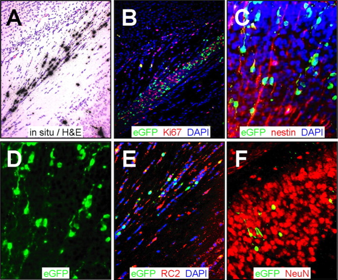
Figure 7.

Virus-infected cells are arrayed as chains, contiguous with radial glia. Paraffin-embedded sections were obtained from the brain 2 d after infection and stained using antibodies against Ki67, nestin, and RC2. Viral infection was evaluated by in situ hybridization (CVB3 5′ untranslated region probe) or by viral protein expression (eGFP). A, In situ hybridization identified infected cells near the retrosplenial cortex in longitudinal arrays. B, Proliferating cells were identified in close proximity to infected cells (eGFP+) in this region. C, Sections stained for nestin and counterstained with DAPI (blue) revealed infected nestin+ cells that appeared to be migratory neuroblasts associated with radial glia. D, Single-channel (eGFP) image of C highlighted the chain-like distribution of infected cells with long extensions entering into the cortex, reminiscent of radial glial cells. E, RC2 staining (red) identified many infected radial glial cells contiguous with the retrosplenial cortex. F, Many infected NeuN+ cells were observed deeper within the cortex. A, B, 20× objective; C, D, 20× objective with an additional approximately threefold computer-generated magnification; E, F, 20× objective with an additional approximately two-fold computer-generated magnification.
