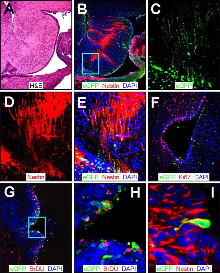
Figure 8.

CVB3 infection adjacent to the fourth ventricle. Sagittal (A-E) and transverse (F-I) paraffin sections of the posterior brain of a newborn mouse at 1 d after infection were immunostained using antibodies against nestin, Ki67, and BrDU. A, Low-magnification H&E-stained sagittal section identified features of the cerebellum, the fourth ventricle, and the ependymal cell layer. B, Infected cells (eGFP+) were evident near the ependymal cell layer in the area below the fourth ventricle and in the region of the dorsal tegmental nucleus. Nestin+ staining (red) was observed stretching across the ependymal cell layer into the surrounding parenchyma. The cyan box represents the area expanded in C-E. C, Higher magnification revealed infected (eGFP+) cells with long cellular processes. D, Similarly, nestin staining demonstrated long extensions of neuronal progenitor cells found near the fourth ventricle. E, Direct colocalization of nestin and infected axonal processes was evident (yellow), indicating that neuronal progenitor cells in the fourth ventricle were susceptible to infection. F, Ki67 staining (red) showed many proliferating cells near the ependymal cell layer and infected regions of the fourth ventricle. G, Similarly, many BrDU cells (red) were found closely associated with infected cells near the ependymal cell layer. H, Higher magnification (G, cyan box) demonstrated direct colocalization of many infected cells with BrDU staining. I, Many nestin+ (red) infected cells protruded through the ependymal cell layer in the fourth ventricle, consistent with infected type B stem cells, as seen near the lateral ventricle (Fig. 4).
