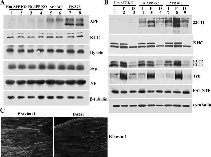
Figure 3.
Expression and anterograde axonal transport of kinesin-1 and Trk receptor are APP independent. A, Protein extracts of sciatic nerves from wild-type mice (APP WT), mice with either homozygous or heterozygous APP depletion (Hm APP KO and Ht APP KO, respectively), and transgenic mice harboring FAD-linked APPswe (Tg2576 mice). Immunoblots for KHC, dynein, synaptophysin (Syp), neurofilament (NF), and β-tubulin show no differences at steady-state levels between the different genotypes, indicating that fast, slow, anterograde, and retrograde axonal transport are not affected by APP expression. B, Protein extracts prepared from ligated sciatic nerve of Ht APP KO, Hm APP KO, and APP WT mice. APP accumulation is evident in the proximal ligature of sciatic nerve of APP WT mice (22C11 panel, lane 8); reduced accumulation is observed in the proximal ligature of Ht APP KO mouse sciatic nerve (22C11 panel, lane 5). APP expression cannot be detected in the sciatic nerve of APP KO mice (22C11 panel, lanes 1-3). The accumulation level of Trk receptor (Trk panel), kinesin heavy chain (H2 antibodies, KHC panel), kinesin light chain (63-90 antibodies, KLC panel), and PS1 (PS1-NTF antibodies, PS1-NTF panel) at the proximal stump of the ligation site is comparable in sciatic nerve of APP-ablated and wild-type nontransgenic mice (compare lanes 2, 5, 8). I, Intact sciatic nerve; P, proximal stump of ligated sciatic nerve; D, distal stump of ligated sciatic nerve. C, Kinesin-1 expression in longitudinal sections of ligated sciatic nerve as detected by immunolabeling using anti-KHC H2 antibodies. Note kinesin-1 accumulation toward the proximal stump of the ligature.

