
Figure 4.
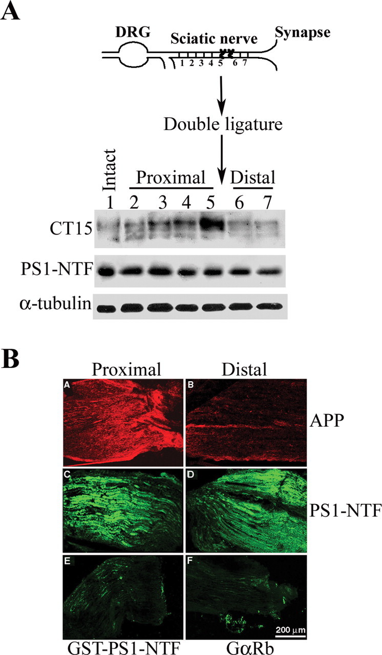
PS1 is not cotransported with APP in the same vesicular compartment by fast axonal transport. A, Top, Schematic diagram of sciatic nerve double ligation and nerve segments examined for protein expression. Bottom, Expression level of APP and PS1 in nerve segments at varying distances from the site of ligation in sciatic nerve of nontransgenic mice. APP expression level increases toward the site of ligation, whereas it decreases away from the distal ligature (CT15 panel, lanes 1-5). In contrast, PS1 expression level is comparable in intact sciatic nerve and nerve segments at varying distances from the site of ligation (PS1-NTF panel, lanes 1-7). B, APP and PS1 immunoreactivity in cryostat sections of the proximal and distal ends of ligated sciatic nerve. A, B, APP is detected using CT15 antibodies. Note the accumulation of APP proximal to the ligature (A), whereas weak immunoreactivity is observed at the distal stump (B). C, D, PS1 immunoreactivity as detected by affinity-purified αPS1-NTF antibodies; C, proximal stump; D, distal stump. Note that neither accumulation at the proximal stump nor reduced immunoreactivity at the distal stump can be detected. E, Specificity of immunostaining for PS1 was confirmed using αPS1-NTF antibodies preincubated with GST-PS1-NTF (see Materials and Methods). F, Immunostaining of mouse sciatic nerve cryostat sections using secondary antibodies only (fluorescein-conjugated goat anti-rabbit). Scale bar, 200 μm.