
Figure 8.
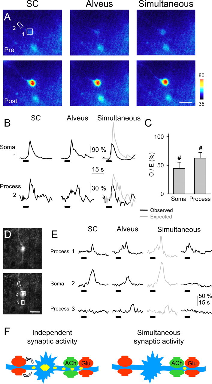
Astrocytic subcellular regions exhibited synaptic-mediated Ca2+ modulation. A, Pseudocolor images representing fluorescence intensities of a fluo-3-filled astrocyte before (Pre) and 10 s after (Post) independent or simultaneous stimulation of the SC and alveus (30 Hz, 5 s). Scale bar, 15 μm. B, Fluorescence intensity changes in the astrocytic soma and a restricted region of the astrocytic process marked with boxes 1 and 2, respectively, in A. C, Ratio between observed and expected Ca2+ elevations in the soma (n = 14) and restricted regions at the processes (n = 24). Significant differences from control values were established by the Student's t test at #p < 0.001.D, Fluorescent images of a fluo-3-filled astrocyte. Scale bar, 15 μm. E, Fluorescence intensity changes in restricted regions of two astrocytic processes (1 and 3) and soma (2) marked with boxes in D, evoked by independent or simultaneous stimulation of the SC and alveus (30 Hz, 5 s). Note that after alveus stimulation, Ca2+ increased in region 1 and later propagated with delay to regions 2 and 3. After simultaneous stimulation, Ca2+ increased in region 1 but failed to propagate to regions 2 and 3. F, Schematic drawing representing a hypothetical consequence of the Ca2+ signal modulation. Under independent high-frequency synaptic activity of either pathway (left), astrocyte Ca2+ elevations initiate in specific processes and then propagate to the soma and other processes, eventually leading to long-distance neuromodulation by Ca2+-dependent release of glutamate (arrows). However, simultaneous high-frequency synaptic activity prevents the intracellular propagation of the astrocyte Ca2+ signal and its long-distance neuromodulatory effects.