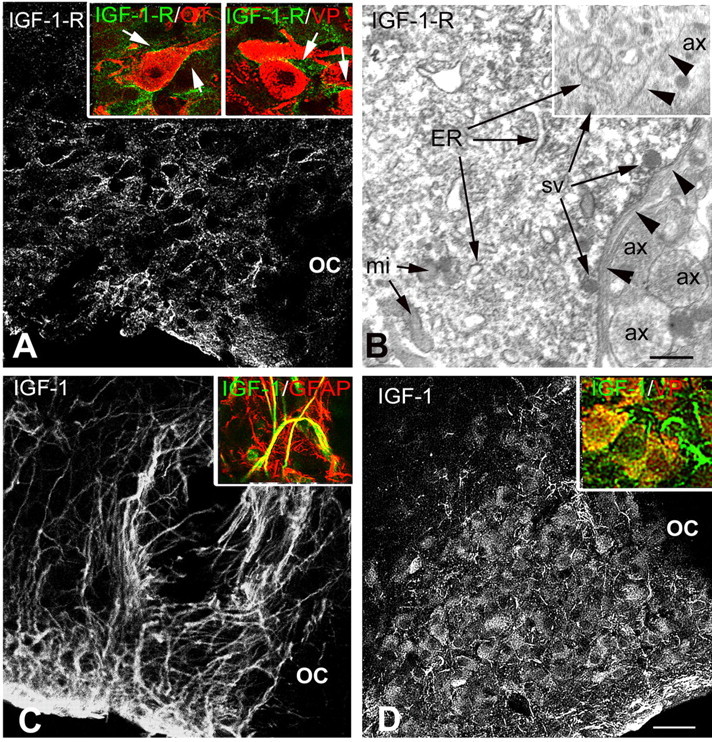
Figure 1.

Immunocytochemical localization of IGF-1 receptor and IGF-1 within the SON. A, At the confocal microscopy level, immunostaining for IGF-1 receptor (IGF-1-R) is localized at the periphery of cell bodies dispersed throughout the SON. Insets, Double immunostaining for IGF-1-R and either OT or VP neurophysins showing that the receptor is localized at the periphery of both types of SON neurons (arrows). B, At the electron microscopy level, IGF-1-R immunostaining appears as electron-dense precipitates associated with both the endoplasmic reticulum and the limiting plasma membrane (arrowheads) of SON neuronal cell bodies, whereas surrounding structures are unlabeled. Inset, No immunostaining is found, regardless of whether associated with the endoplasmic reticulum (ER) or plasma membrane (arrowheads), in a control section processed in the absence of the primary antibody. C, Immunostaining for IGF-1 is associated with elongated astrocytic-like processes in a control untreated rat, where it colocalizes with GFAP (inset). D, In a colchicine-treated rat, IGF-1 immunostaining is detected within most SON neurons and both VP-labeled neurons (inset, yellow) and VP-negative processes (inset, arrow). ax, Axon; mi, mitochondria; OC, optic chiasma; sv, secretory vesicle. Scale bars: A, C, D, 50 μm; insets, 20 μm; B, 0.5 μm.
