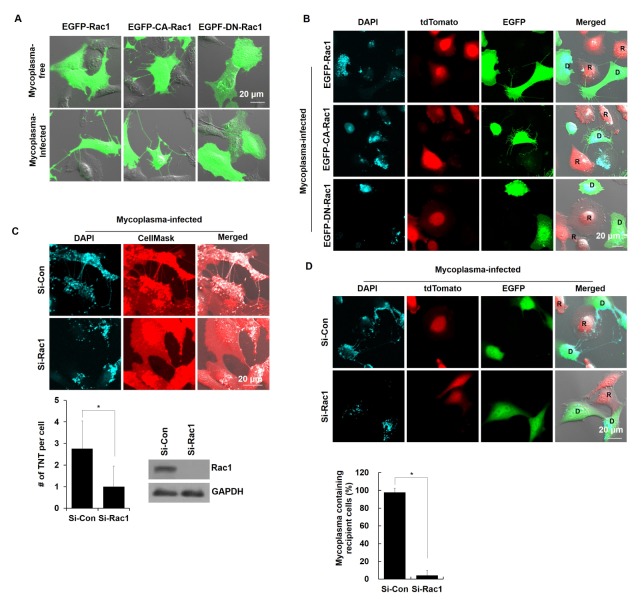
Fig. 4.
Genetic inhibition of Rac1 prevented the cell-to-cell dissemination of M. hyorhinis via TNTs. (A) Mycoplasma-free and -infected NIH3T3 cells were overexpressed with an EGFP-Rac1, EGFP-Rac1-Q61L (constitutively active form of Rac1, EGFP-CA-Rac1), and EGFP-Rac1-T17N (dominant negative form of Rac1, EGFP-DN- Rac1) and their TNTs were observed in the live state. Boxed regions are enlarged in the right panels. (B) M. hyorhinis-free or -infected NIH3T3 cells were overexpressed with EGFP-Rac1, EGFP-CA-Rac1 and EGFP-DN-Rac1 and co-cultured with tdTomato-expressing Chang Liver cells in a mixed co-culture system. After 24 h, the cells were stained with DAPI (3 μM) in the live state. D and R represent the donor and recipient cells, respectively. (C) M. hyorhinis-infected NIH3T3 cells were transfected with scrambled siRNA (si-Con) or si-Rac1 for 24 h and stained with DAPI (3 μM) and CellMask™ (5 μg/ml) in the live state. Boxed regions are enlarged in the bottom panels. The number of TNTs per cell was counted and is represented as the mean ± s.d. *P < 0.01. The expression level of Rac1 and GAPDH was determined by immunoblotting. (D) M. hyorhinis-infected EGFP-expressing NIH3T3 cells (donor cells) were transfected with scrambled si-Con or si-Rac1 for 24 h and co-cultured with tdTomato-expressing cells (recipient cells) in a mixed co-culture system. After 24 h of co-culture, the cells were stained with DAPI (3 μM) in the live state. The percentage of DAPI-stained cells among all recipient cells was statistically determined and is represented as the mean ± s.d. *P < 0.01. One hundred cells were observed in ten different fields under each condition.

