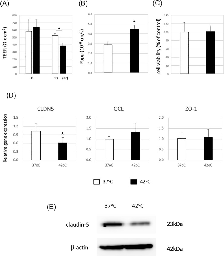
Fig 2. The barrier function of iPS cell-derived brain microvascular endothelial cells is disrupted under heat stress.
iPS cell-derived brain microvascular endothelial cells on Transwell membranes were incubated at 37°C or 42°C for 12 h. (A) TEER values were monitored before and after heat stress exposure. Data are represented as the mean +/- S.D. from three independent experiments (n = 3). *P<0.05 (two-tailed Student’s t-test; compared with 37°C). (B) NaF flux across the cell monolayer was measured. Data are the mean +/- S.D. from three independent experiments (n = 3). *P<0.05 (two-tailed Student’s t-test; compared with 37°C). (C) Cell viability was measured by alamarBlue. Data are the mean +/- S.D. from three independent experiments (n = 3). *P<0.05 (two-tailed Student’s t-test; compared with 37°C). (D) Gene expression levels of claudin-5, occludin, and ZO-1 were measured by quantitative RT-PCR. Gene expression levels of cells at 37°C were set to 1.0. Data are the mean +/- S.D. from three independent experiments (n = 3). *P<0.05 (two-tailed Student’s t-test; compared with 37°C). (E) Expression levels of claudin-5 were evaluated by western blotting. One representative image from three independent experiments is shown.

