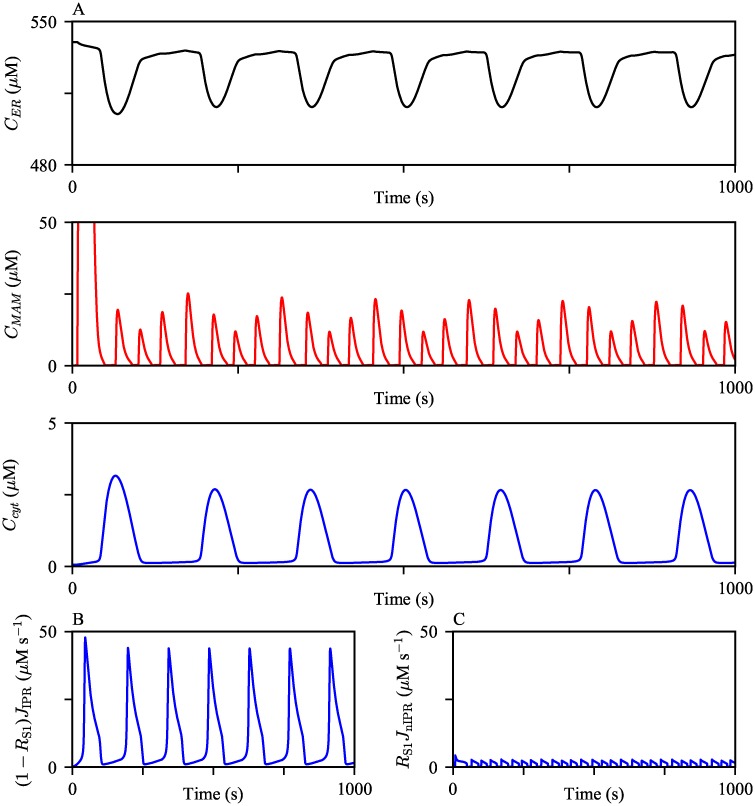
Fig 4. Ca2+ oscillations generated from the model exhibit varying orders of magnitude in different compartments.
(A) The model was given continuous stimulation of IP3 with Ps = 0.3 μM. From the top, the panels show Ca2+ oscillations in the ER, the MAM, and the bulk cytosol. (B and C) The magnitudes of IPR Ca2+ fluxes from the ER to the bulk cytosol and the MAM, respectively, during the oscillations shown in (A).

