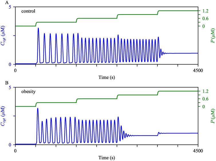
Fig 13. Robustness of Ca2+ oscillations under different model conditions.
We perturbed (A) the control model and (B) the obesity model with gradually increasing stimulation. Initially, Ps was at 0 μM, then was increased to 0.3 μM, 0.6 μM, 0.9 μM, and then to 1.2 μM at t = 500 s, 1500 s, 2500 s, and 3500 s, respectively. The cytosolic Ca2+ concentrations are shown in blue, with the scale on the left y-axis. The green timeseries represent the IP3 concentration, with the scale on the right y-axis.

