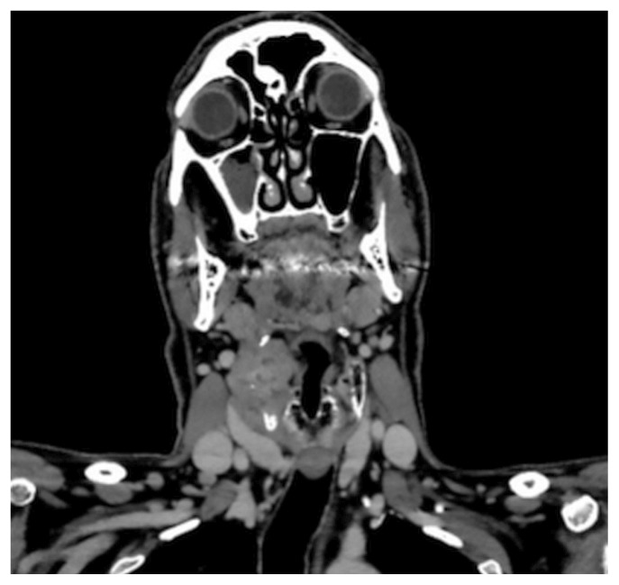
FIGURE 1.
Computed tomography imaging of the patient’s neck. A coronal slice shows a 5.0×3.8×3.8 cm mass centred on the right thyroid cartilage and invading the right paraglottic space and the right true vocal cord, and appearing inseparable from the right strap muscles, together with prominent right cervical lymph nodes.

