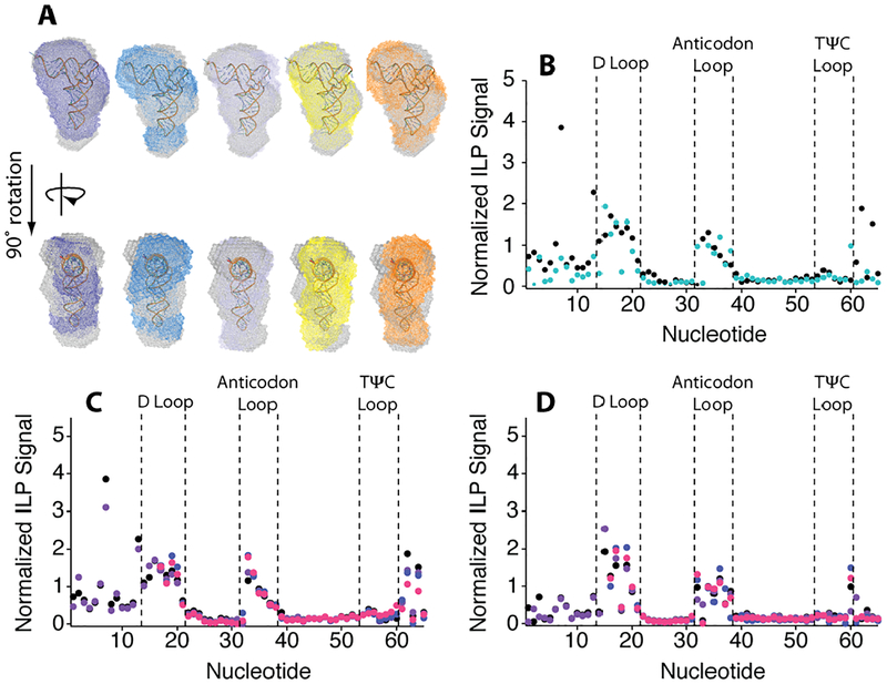
Figure 3.
Small angle X-ray scattering bead models of M1–M5 aligned with the WT bead model and the tRNA crystal structure in buffer with 2.0 mM Mg2+. (A) Alignment of WT (grey) and M1 (purple), M2 (blue), M3 (light blue), M4 (yellow), and M5 (orange) DAMAVER envelopes. (B) Normalized ILP signal comparing WT (black) with M5 (teal) in buffer. Normalized ILP Signal of (C) WT and (D) M5 normalized ILP signal in (black) buffer, (purple) 20% PEG8000, (blue) Mg2+-chelated amino acids and (pink) 20% PEG8000 with Mg2+-chelated amino acids in the background of 2.0 mM free Mg2+. Nucleotides 1–15 were not analyzed in samples containing Mg2+-chelated amino acids due to salt contamination.

