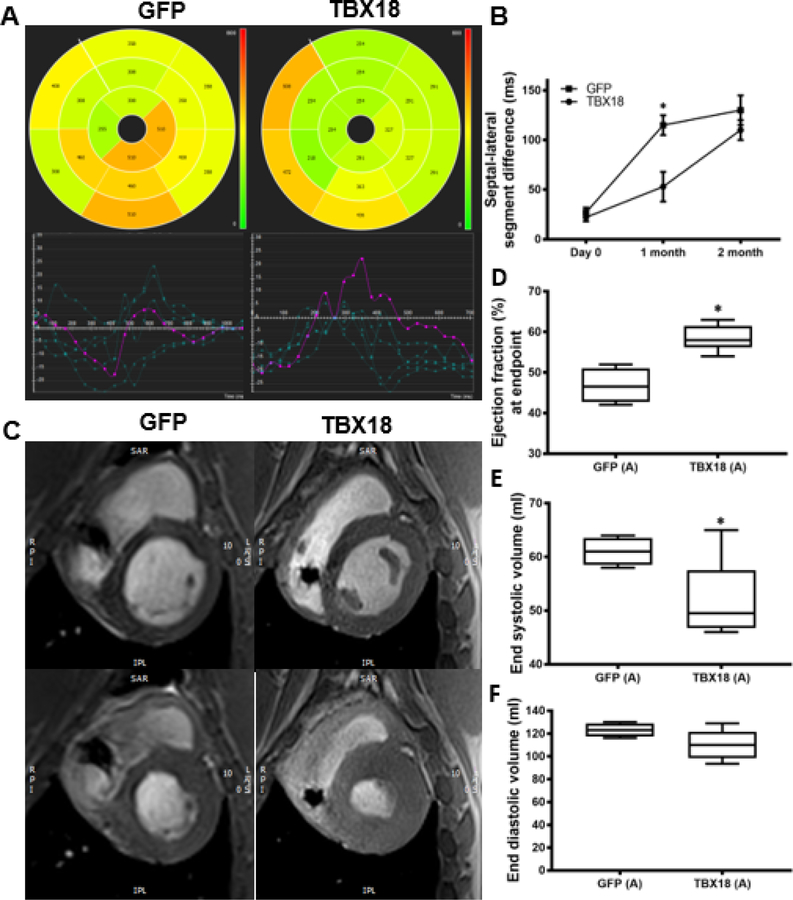
Figure 2: Group A – Early intervention protocol: changes in mechanical dyssynchrony, and, and left ventricular systolic function in TBX18 antegrade biological pacemaker treated-animals compared to controls.
(A) 2D polar maps of radial strain with AHA segmentation representing time to peak of each segment with plots (below) depicting the time to peak radial strain of individual chords (cvi42®). (B) Septal-lateral segment difference was better in TBX18 pigs one month following AV block (115±10ms GFP, 53±15ms TBX18, P=0.001) (C) Representative post processed MRI (cvi42®) identifying end diastolic (top) and end systolic (bottom) phases (D) Endpoint ejection fraction was higher in TBX18 animals (46.75±2% GFP, 58.5±1.3% TBX18, p=0.001). (E-F) Additionally, TBX18 pigs demonstrated lower end systolic (61.1±1.2ml GFP, 52±3ml TBX18 p=0.04) and end diastolic volume (123±3ml GFP, 110±5ml TBX18, p=0.1)

