
Fig. 3. Effects of acupuncture on hypothalamic ARC neuron activities.
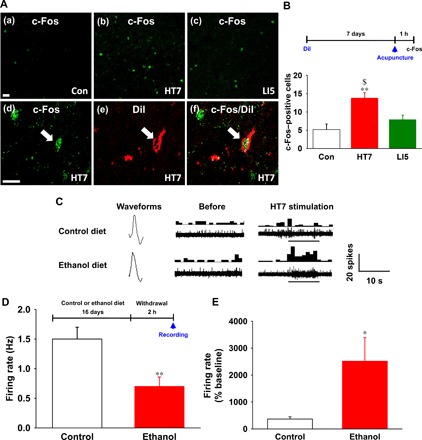
(A and B) Immunohistochemical staining of c-Fos in ARC neurons of control rats (a) and rats given acupuncture at HT7 or LI5 (b and c). A significant increase in the number of c-Fos–positive cells in the ARC was shown in rats subjected to HT7 acupuncture compared to control rats or rats subjected to LI5 acupuncture [#P < 0.05 versus Con, n = 6 per group; (B)]. c-Fos–positive cells (d) in ARC neurons labeled with DiI (e) in HT7 acupuncture–treated rats (f). Scale bars, 50 μm (200×). (C) Typical waveforms of spontaneous and evoked activity of ARC neurons before and during HT7 acupuncture in control (n = 7) or ethanol (n = 7) diet rats. Left, single action potential; middle and right, the continuous single action potential of real-time (waveform) (bottom) and peri-stimulus time histogram (one bin width per second, top). (D) Basal firing rates in the control and ethanol diet rats before acupuncture stimulation. **P < 0.01 versus control group. (E) Effect of acupuncture on firing rate of control and ethanol diet rats. Single-cell activity was analyzed by graphic recording for 20 s at rest and 10 s during acupuncture treatment. *P < 0.05 versus control group. Graphs represent mean ± SEM (n = 7 per group).