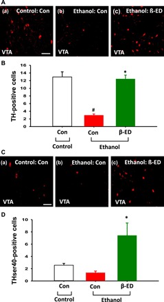
Fig. 6. Effects of intra-NAc infusions of β-endorphin on TH expression and phosphorylation in the VTA.

(A and B) Immunohistochemical staining of TH in VTA neurons of the ethanol group given intra-NAc infusions of β-endorphin at 2 hours after ethanol withdrawal (c). Another group of control diet rats (a) (Con-control group, n = 7) and ethanol diet rats (b) (Con-ethanol group, n = 7) received artificial cerebrospinal fluid infusion in place of β-endorphin infusion. A significant increase in the number of TH-positive cells in the VTA was shown in rats (β-ED-ethanol group, n = 6) subjected to intra-NAc infusions of β-endorphin compared to control rats [#P < 0.05, Con-control versus Con-ethanol; *P < 0.05, Con-ethanol versus β-ED-ethanol; (B)]. Scale bar, 50 μm (200×). (C and D) Immunohistochemical staining of THser40, the phosphorylation form of TH, in VTA neurons of ethanol group given intra-NAc infusions of β-endorphin at 2 hours after ethanol withdrawal (c). Another group of control diet rats (a) (Con-control group, n = 7) and ethanol diet rats (b) (Con-ethanol group, n = 7) received artificial cerebrospinal fluid infusion in place of β-endorphin infusion. A significant increase in the number of THser40-positive cells was shown in the VTA of rats (β-ED-ethanol group, n = 6) subjected to intra-NAc infusions of β-endorphin compared to control rats [*P < 0.05, Con-ethanol versus β-ED-ethanol; (E)]. Scale bar, 50 μm (200×).
