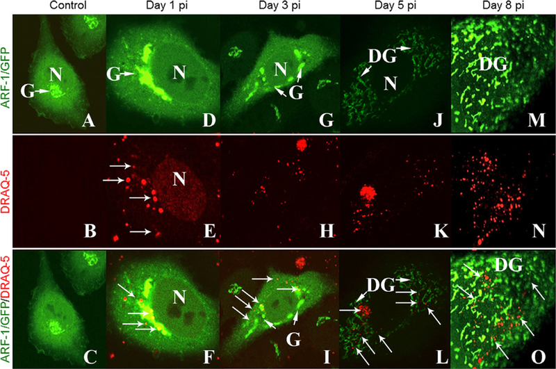
Figure 2.

Live imaging of microsporidia and Golgi Reorganization. Plated HeLa cells were transfected with Arf1-GFP DNA plasmid before inoculating the cultures with DRAQ5 labeled Anncaliia algerae, and imaged at different time points during infection. A-C. ControlHeLa cells (No infection) showing the distribution of Arf-1, in the Golgi compartment. Note its perinuclear location. D-F. Day 1 postinfection, Arf-1 distribution is similar to the control cells and there are organisms (arrows) in the cells. G-I. On the third day PI, the Golgi fragments and shows a perinuclear distribution. Parasites are visible near the host nucleus and Golgi. J-L. Day 5 PI, the Golgi is disorganized and fragmented into mini-Golgi like structures that are redistributed throughout the cytosol coinciding with the parasite distribution. M-O. By the eighth day, the new mini-Golgi structures are clearly visible allover the host cell cytosol and are associated with parasite progeny (arrows). Arrows = DRAQ5 stained parasite nuclei; DG = mini-Golgi; G = Golgi; N = host nuclei.
