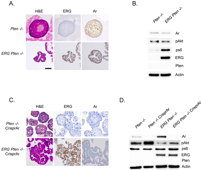
Figure 2. Establishment and characterization of Pten−/− and ERG Pten−/− prostate cancer organoids.
A) Tumors derived from our Ptenlox/lox and ERG Ptenlox/lox GEM models were used to establish prostate cancer organoids (3 clones for each genotype) and characterized in 3-D culture conditions for histology, and immunohistochemistry was performed for ERG and AR. B) Western blotting confirming loss of Pten, activation of PI3K pathway, and ERG over-expression. C) Pten−/− and ERG Pten−/− organoids underwent AR Crispr (3 individual clones for each genotype) and were characterized in 3-D culture conditions for histology, and immunohistochemistry was performed for ERG and AR. D) Western blotting confirming loss of Pten, activation of PI3K pathway, ERG over-expression, and loss of AR following AR Crispr.

