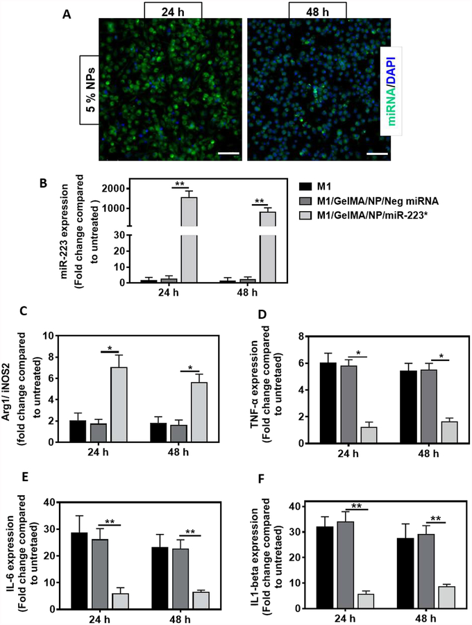
Figure 4. miRNA transfection and macrophage re-polarization studies in J774A.1 macrophages using GelMA/NP/miR-223* hydrogels in a trans-well set up.
(A) Representative fluorescent microscope images for transfection of Cy3-labeled miRNA released from GelMA/NP/ miRNA hydrogel, at the concentration of 5.0 %(w/w), in J774A.1 macrophages on 24, and 48 h post-incubation. Green color represents Cy3-labeled miRNA and blue color represents cell nuclei, respectively (Scale bar = 200 μm). (B) miR-223 expression was quantified by miR-223 specific Taqman™ assay performed in J774A.1 macrophages on 24, and 48 h post-incubation with GelMA/NP/Neg miRNA, and GelMA/NP/miR-223* hydrogels. The M1 represents cells that received LPS+IFN-Ƴ treatment for 16 h before incubation with either groups. Neg miRNA represents negative control miRNA and miR-223* represents miR-223 5p mimic. The gene expression level was normalized to untreated cells that did not receive any treatment. U6 snRNA was used as endogenous housekeeping gene. (C) In vitro macrophage re-polarization. qPCR analysis of Arg-1 (M2 marker)/iNOS-2 (M1 marker) gene expression was conducted on 24, and 48 h post-incubation with GelMA/NP/Neg miRNA, or GelMA/NP/miR-223* hydrogels. β-actin was used as a house keeping gene. (D-F) Anti-inflammatory effects of miR-223* transfection in J774A.1 macrophages. Decreased expression of pro-inflammatory cytokines; TNF-α, IL-6, and IL1-β mRNA levels was observed upon 24 and 48 h post-incubation with GelMA/NP/Neg miRNA and GelMA/NP/miR-223* hydrogels. The gene expression level was normalized to untreated cells that did not receive any treatment. β-actin was used as a house keeping gene. *p<0.05. Data are represented as mean ± SD (. *p<0.05, and **p < 0.01, compared to GelMA/NP/Neg miRNA group with n =4 individual experiments per group).

