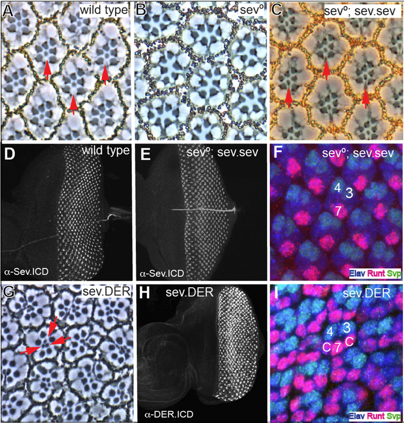
Figure 1. Rescue of sev° by Sev expression, and the effects of DER expressed at Sev levels.
(A) Phase contrast image through a wild type retina in which the rhabdomeres of six outer photoreceptors are arrayed in an asymmetric trapezoid shape. In the center of the array lies the small central rhabdomere of R7 (red arrows). (B) R7 is absent from every ommatidium in sev° eyes. (C) sev expressed under sev transcriptional control (sev.sev) restores R7s (red arrows) to all sev° ommatidia. (D) An antibody raised against the C-terminus of Sev highlights the typical expression pattern in wild type eye discs. (E) α-Sev.ICD staining of sev°; sev.sev eye discs show the normal Sev expression pattern. (F) In sev°; sev.sev eye discs, normal pattern formation is evident with R7 specified at the correct time and place. At the two-cone-cell-stage, R7s (highlighted by Elav (blue) and Runt (red) expression) are seen on the other side of the ommatidium from R3/4 labeled by Elav and Svp (green). (G) In sev.DER adult eyes, many R7-like cells (red arrows) are evident in each ommatidiaatidium. (H) A sev.DER eye disc labeled with an antibody raised against the C-terminal region of DER. Strong DER staining is observed in the Sev-expression cells superimposed upon the normal DER expression pattern (see Fig.2D). (I) At the two-cone-cell-stage, sev.DER eye discs show cells in cone cell positions (c) differentiating as R7-like photoreceptors (expressing Runt and Elav).

