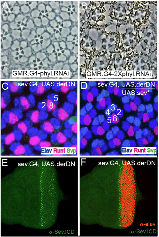
Figure 7. Selective R7 sensitivity to phyl gene function reduction, and Sev* rescue of R3/4 DER function.
(A) Section through a GMR.GaU; UAS.phyl adult eye shows the typical sev° phenotype with each ommatidium specifically lacking R7. (B) When phyl gene function is further compromised (GMR.GaU; 2XUAS.phyl.RNAi) only 4 large rhabdomere photoreceptors are evident in most ommatidia. (C) sev.Gal4; UAS.derDN eye discs stained for Elav (blue) Svp (green) and Runt (red). Only R2/8/5 clusters are evident indicating that photoreceptor recruitment ceases at this stage. (D) When UAS.sev* is introduced into the sev.Gal4; UAS.derDN background, the recruitment of the R3/4 photoreceptors is rescued. (E) α-Sev staining (green) indicates normal expression in R3/4 precursors in the early ommatidia, but is degenerate in later ommatidiaatidia. (F) Co-staining of sev.Gal4; UAS.derDN with Sev and Elav highlights the normal R3/4 Sev expression before photoreceptor differentiation begins.

