
Figure 3. Oncogenic KRAS promotes EMT and acquisition of CSC phenotypes in gastric epithelial cells.
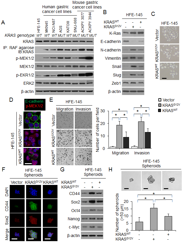
(A) Western blot for RTK-RAS pathway proteins in gastric epithelial cells and human and mouse gastric cancer cell lines. The presence (MUT) or absence (WT) of an oncogenic KRAS mutation is denoted. (B) Western blot for EMT-related proteins in HFE-145 cells transduced with oncogenic KRAS (KRASG12V), wild-type KRAS (KRASWT) or control (Vector). (C) Morphology of HFE-145 in tissue culture following transduction with KRASG12D, KRASWT, or Vector. Scale bar 10 μm. (D) Confocal photos following immunofluorescent staining of HFE-145 cells transduced with KRASG12D, KRASWT, or Vector for E-cadherin (green) and p-MEK1/2 (red). Scale bar, 20 μm. (E) Migration and invasion assays for HFE-145 cells transduced with KRASG12D, KRASWT, or Vector as determined by transwell assay. (F) Immunofluorescence of HFE-145 spheroids for DAPI (blue), CD44 (green), and Sox2 (red) following transduction with KRASG12D, KRASWT, or Vector. Scale bar 50 μm. (G) Western blot for self-renewal proteins in HFE-145 cells transduced with KRASG12D, KRASWT, or Vector. (H) Photos and graph of HFE-145 cells following transduction of KRASG12D, KRASWT, or Vector and grown in spheroid formation conditions. Scale bar 50 μm. Bars represent standard deviation. *p<0.05.