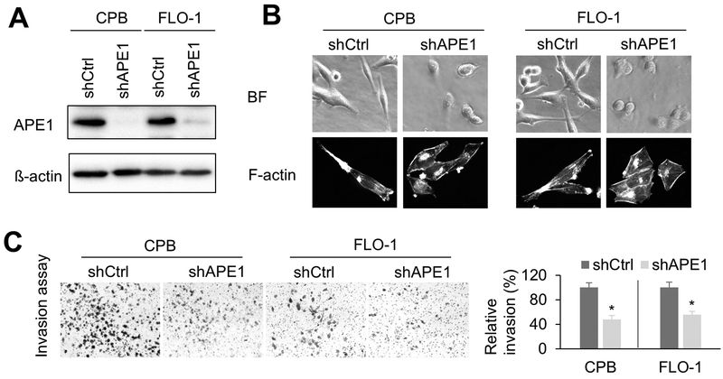
Figure1. silencing decreases invasion capacity.
A, Western blot analysis of CPB and FLO-1 cells with APE1-knockdown (shAPE1) or control (shCtrl). B, Representative cell images of bright field (BF) and F-actin staining in APE1-knockdown cells or control cells. Alexa Fluor™ 488 Phalloidin was used for F-actin staining. C, invasion assays were performed using APE1-knockdown cells or respective control cells. The results were expressed as the mean ± SEM of three independent experiments. *, p < 0.05.

