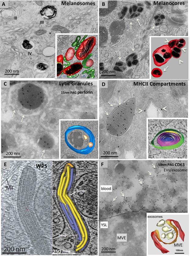
Figure 1. Examples of ELRO ultrastructure.
Conventional electron microscopy (A, B), immunogold labelling on ultrathin cryosections (C, D, F), cryomicroscopy (E) and three-dimensional (3D) reconstructions of electron tomograms of model ELROs and their derivatives (A, B, C, D, E, F). A, stage II immature and stage III and IV mature melanosomes in an MNT-1 human melanoma cell fixed by high pressure freezing and embedded in plastic by freeze substitution. Note the striated appearance in stages II and III. Inset: 3D reconstruction showing dark melanin on fibrillar structures (black) in stage III/ IV melanosomes. Unpigmented fibrillar structures present in stage II melanosomes are shown in white. Melanosomes are surrounded by tubular membranes (green), corresponding to endosomal transport carriers.Ribosomes are in blue B, melanocore containing organelles (arrows) in a keratinocyte in human skin biopsies. Inset: 3D reconstruction. Note several melanocores (black) enclosed by a single membrane (red). These organelles appear isolated (arrow) or in a network of clusters (arrowheads). C, lytic granules (arrows) of a human cytotoxic T cell depicting the characteristic dense core containing perforin, immunolabelled with 15 nm protein A gold particles (PAG), surrounded by small membrane vesicles. Inset: 3D reconstruction showing the limiting membrane (blue) and ILVs (yellow). The dense core is not pseudocoloured. D, an ultrathin cryosection of a mouse dendritic cell showing MHC class II (MHCII) compartments (arrows) immunolabeled for MHCII molecules with 10 nm PAG and endosomes (arrowheads) immunolabeled for the endosomal protein EEA1 with 15 nm PAG. Inset: 3D reconstruction showing the multiple concentric membrane layers of an MHCII compartment. E, Cigar-shaped WPBs in a thick section of a human umbilical vein endothelial cell visualized by cryomicroscopy (left panel). The right panel shows a 3D reconstruction of the vWF tubules (blue and yellow) contained within the WPB. F, ultrathin cryosection of a zebrafish embryo expressing CD63-pHluorin in the yolk syncytial layer and labelled for GFP with 10 nm PAG. The plasma membrane of the yolk sac layer is indicated by the dashed line. Note the MVE in the yolk cell and numerous membrane vesicles labelled for CD63 (arrows) in the blood. Inset: 3D reconstruction of an MVE in a HeLa cell in the process of fusing with the plasma membrane. Magnifications are indicated in the panels.

