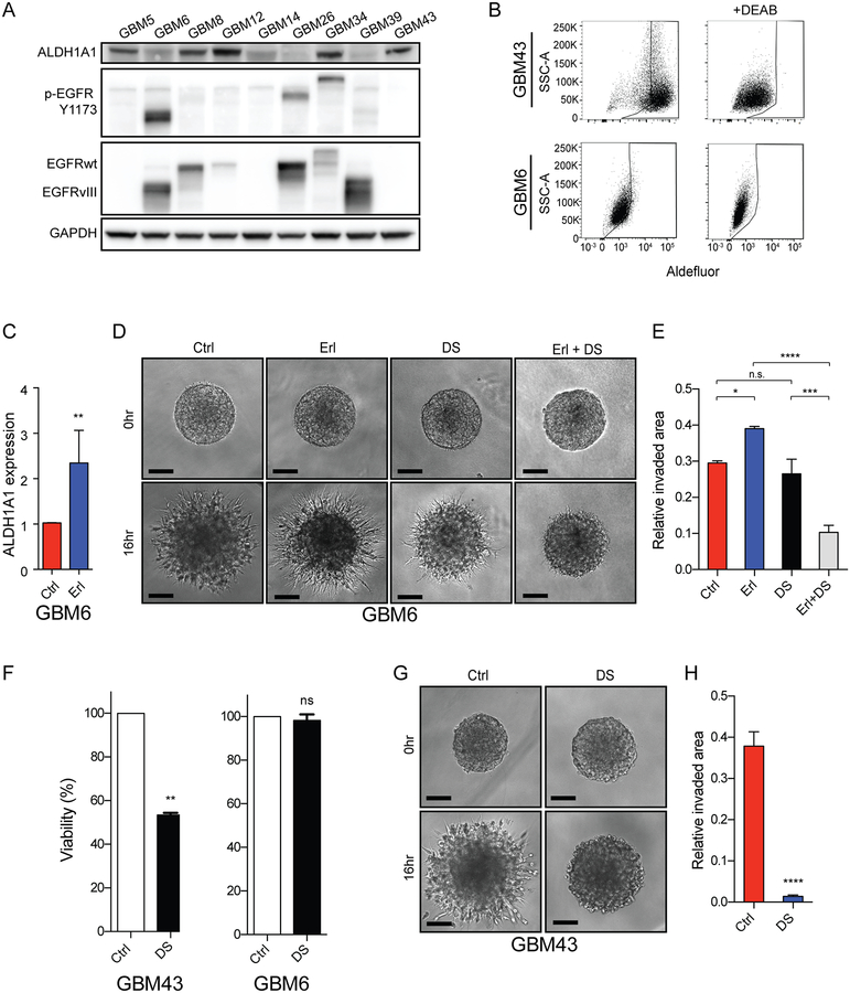
Figure 5. Induction of ALDH1A1 expression and a pro-invasive phenotype upon EGFR inhibition in an EGFR-activated patient-derived GBM xenograft.
(A) ALDH1A1 expression and EGFR expression and phosphorylation across a cohort of human GBM xenografts. GAPDH protein loading control. (B) Aldefluor activity (left panels) in GBM43 (top) and GBM6 (bottom) relative to baseline fluorescence established by inhibiting ALDH with DEAB (right panels). (C) Erlotinib-induced increase in ALDH1A1 mRNA expression in GBM6. (D-E) Erlotinib-induced and disulfiram-sensitive increase in invasion in GBM6. (F) ALDH1A1-high GBM43 demonstrate increased sensitivity to disulfiram treatment than GBM6 and (G-H) GBM43 invasion is inhibited by disulfiram treatment. Representative images and quantification, mean ± SEM of biologic triplicate. *p<0.05; **p<0.01; ***p<0.0005; ****p<0.0001.

