
Figure 2.
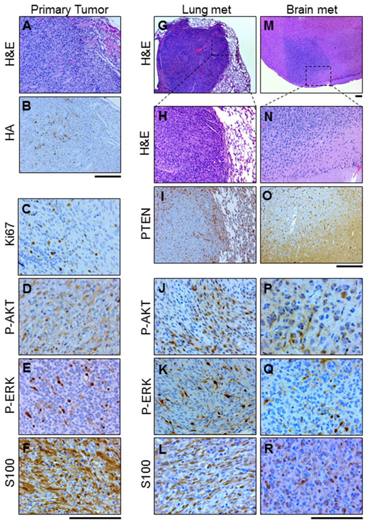
Histological analysis of the primary tumor and metastatic tumor tissue show similarities with human melanoma. Representative images of serial sections from a primary tumor, lung lesion, and brain lesion in BRAFV600E;Cdkn2a−/−;Pten−/−;AKT1E17K mice are shown. A, primary tumor stained with H&E. B, IHC for HA performed on the primary tumor tissue revealed AKT1E17K- expression as well as the presence of C, Ki67 D, P-AKT (S473) E, P-ERK (T202/Y204) and F, S100. G-H, H&E staining was used to identify lung metastases. I, IHC for PTEN revealed expression in normal tissue but absence of expression in lung metastases and the presence of J, P-AKT (S473) K, P-ERK (T202/Y204), and L, S100. M-N, H&E staining was used to identify brain metastases. These tumors were subject to identical IHC analyses as the lung metastases. Analyses revealed the absence of O, PTEN and presence of P, P-AKT (S473) Q, P-ERK (T202/Y204), and R, S100. Scale bars represent 100 μm.