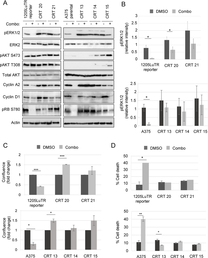
Fig 2. Cell lines generated from combination-resistant tumors displayed ERK½ reactivation.
(A) Western blots of lysates from matched parental cell lines: 1205LuTR reporter for CRT 20 and 21; and A375 for CRT 13, 14, and 15 were blotted for signaling proteins and cell cycle progression markers as indicated (n=3). (B) Quantification of pERK½ intensity normalized to loading control in parental cells and CRTs. Shown are the mean and SEM of five independent experiments, *p<0.05. (C) Parental cells (1205LuTR and A375) and CRTs (20, 21, 13, 14, and 15) were grown for 72 hours in either vehicle (DMSO) or 1 μM PLX4720 and 35 nM PD0325901 and then assayed for 2D growth by measuring live cell counts. Shown are the mean and SEM of three independent experiments, *p<0.05, ***p<0.001 (n=3). (D) Parental cells and CRTs were grown for 72 hours in either vehicle (DMSO) or 1 μM PLX4720 and 35 nM PD0325901 and then processed for annexin V and PI staining as markers for early and late apoptosis. *p<0.05, **p<0.01 (n=3).

