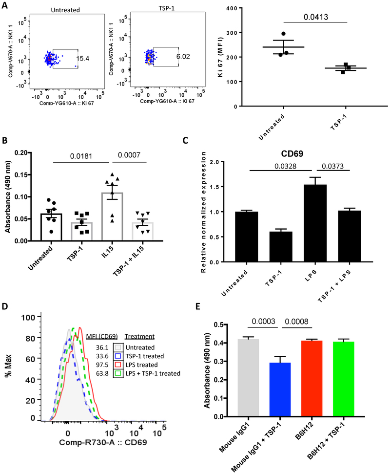
Figure 2: Thrombospondin-1 inhibits NK cell proliferation and activation.
NK cells were isolated from spleens of WT mice by MACS NK isolation kit. (A) Cells were cultured in serum free RPMI for 24 hours with/out thrombospondin-1 (2 nM) followed by fixation, permeabilization and i.c. staining for Ki-67, n = 3 biological replicates, error bars indicate standard error of the mean (SEM). (B) NK cells were cultured in serum free RPMI with/without IL-15 (10 ng/ml) and MTS absorbance was measured after 24 hrs of culture in the presence or absence of thrombospondin-1 (2 nM), n = 7 biological replicates, error bars indicate SEM. (C) Isolated NK cells were divided into 4 groups and cultured in serum free RPMI with thrombospondin-1 (2 nM), LPS (1 μg/ml) or both. mRNA abundance (relative to β-Actin) and (D) cell surface protein levels of CD69 were measured by RT-qPCR (LPS versus TSP-1+LPS treatments, P = 0.0373, n = 3 biological replicates, error bars indicate SEM) and flow cytometry (representative), respectively. (E) Human NK-92 cells were cultured in serum free RPMI with 100 IU IL-2 and treated either with mouse IgG (1 ug/ml) or B6H12 (1 ug/ml) in the presence or absence of human thrombospondin-1 (2 nM) and MTS absorbance was measured after 48 hrs, n = 5 technical replicates, error bars indicate SEM.

