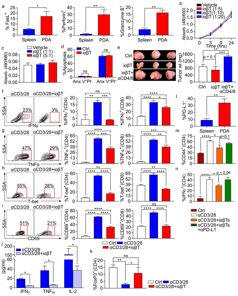
Figure 4. iαβTs induce T cell dependent tumor immunity but are directly suppressive to conventional T cells.
(a) WT mice bearing orthotopic KPC tumors were sacrificed on day 21. Splenic and PDA-infiltrating iαβTs were tested for expression of FasL, Perforin, and Granzyme B (n=5 mice). (b, c) iαβTs were harvested by FACS from orthotopic PDA tumors and cultured in various ratio with KPC tumor cells. (b) Proliferation of KPC tumors cells was tested using the XTT assay. (c) Cytotoxicity against KPC tumor cells was determined in an LDH release assay. (d) iαβTs were harvested by FACS from orthotopic PDA tumors and cultured in 1:1 ratio with KPC tumor cells. KPC tumor cell apoptosis was determined by co-staining for Annexin V and PI. (e) WT mice were orthotopically administered KPC tumor cells admixed with iαβTs. Cohorts were either serially depleted of both CD4+ and CD8+ T cells or administered isotype control before sacrifice on day 21. Representative images and quantitative analysis of tumor weights are shown (n=5/group). This experiment was repeated twice. (f-i) Polyclonal splenic CD4+ or CD8+ T cells were cultured without stimulation, stimulated by CD3/CD28 co-ligation, or stimulated by CD3/CD28 co-ligation in co-culture with iαβTs. CD4+ and CD8+ T cell activation were determined at 72h by their expression of (f) IFNγ, (g) TNFα, (h) T-bet, and (i) CD69. Representative contour plots and quantitative data are shown. (j) Polyclonal splenic CD3+ T cells were stimulated by CD3/CD28 co-ligation, either alone or in co-culture with iαβTs. Cell culture supernatant was tested for expression of IFNγ, TNFα, and IL-2 at 72h. (k) Polyclonal splenic CD4+ T cells were cultured without stimulation, stimulated by CD3/CD28 co-ligation, or stimulated by CD3/CD28 co-ligation in co-culture with iαβTs. CD4+ T cells were tested for expression of FoxP3 at 72h. (l) Spleen and PDA-infiltrating iαβTs were tested for expression of PD-L1 by flow cytometry. (m, n) Polyclonal splenic CD4+ T cells from PD-L1–/– mice were cultured without stimulation, stimulated by CD3/CD28 co-ligation, or stimulated by CD3/CD28 co-ligation in co-culture with WT iαβTs, either alone or with an αPD-L1 neutralizing mAb. CD4+ T cells were tested for expression of (m) CD44 and (n) IFNγ at 72h. Experiments were performed in replicates of 5 and repeated at least 4 times (*p<0.05, **p<0.01, ***p<0.001, ****p<0.0001).

