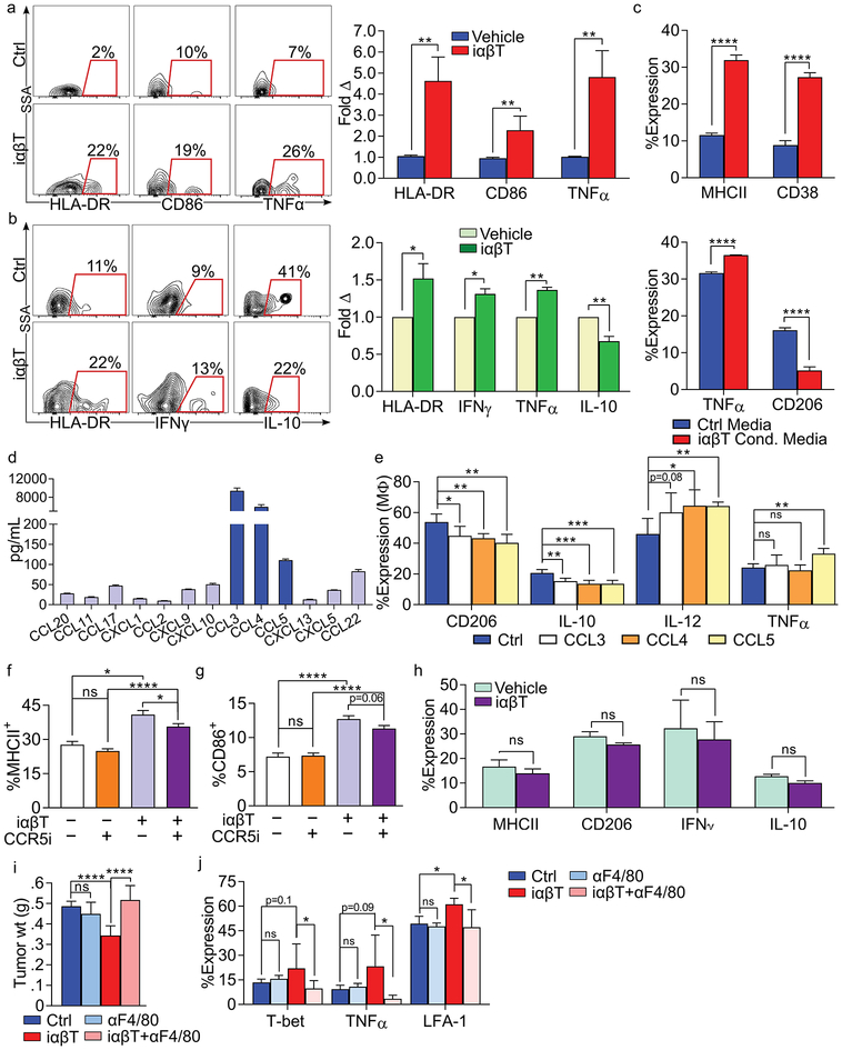
Figure 6. iαβTs induce immunogenic macrophage programming via CCR5 activation.
(a) iαβTs were FACS-sorted from 3 healthy volunteers and co-cultured with autologous naïve PBMC-derived macrophages. At 24h, macrophage expression of HLA-DR, CD86, and TNF-α were determined by flow cytometry. Experiments for each individual were performed in triplicate. Representative contour plots are shown and quantitative data is presented as fold-change in expression compared with macrophages cultured alone. (b) Human PDOTS were treated with autologous iαβTs or vehicle. Tumor-associated macrophages were tested for expression of HLA-DR, IFNγ, TNFα, and IL-10. Representative contour plots are shown and quantitative data is indicated as fold-change in expression compared to vehicle treatment (n=5 patients). (c) Naive macrophages were cultured with iαβT cell conditioned media or control media. After 24h, macrophage expression of MHCII, CD38, TNF-α, and CD206 were determined by flow cytometry. This experiment was repeated 3 times. (d) iαβTs were FACS-sorted from orthotopic PDA tumors and cultured for 24h. Cell culture supernatant was analyzed in a chemokine array. This experiment was repeated twice (n=5). (e) WT bone marrow-derived macrophages were treated with rCCL3, rCCL4, rCCL5, or vehicle and assessed at 24h. (f, g) WT derived macrophages were cultured alone or co-cultured with PDA-infiltrating iαβTs for 24h. A CCR5 small molecule inhibitor (CCR5i) or vehicle was added to select wells. Macrophage expression of (f) MHC II and (g) CD86. This experiment was repeated 3 times (n=5/group). (h) CCR5–/– macrophages were cultured with PDA-infiltrating iαβTs for 24h. Macrophage expression of MHC II, CD206, IFNγ, and IL-10 were determined. This experiment was repeated 4 times in replicates of 5. (i, j) Orthotopic PDA tumors were harvested from control WT mice or WT mice treated with αF4/80, iαβT cell transfer, or αF4/80 plus iαβT cell transfer. (i) tumor weights were measured and (j) CD8+ T cell activation was determined by expression of T-bet, TNFα, and LFA-1 (n=7/group; *p<0.05, **p<0.01, ***p<0.001, ****p<0.0001).

