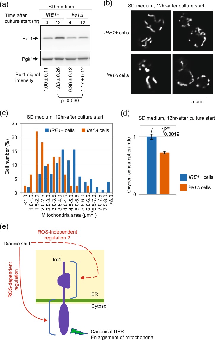
Figure 8.
Ire1-dependent enhancement of respiration and mitochondria size after diauxic shift. IRE1+ cells and ire1Δ cells were cultured in SD medium for the indicated durations. (a) Cell lysates (equivalent to 0.22 OD600 cells) were analyzed by anti-Por1 Western blotting. Anti-Pgk1 Western blot serves as a loading control. See Fig. S12 for the uncropped blot image. (b,c) Cells producing mitochondria-located GFP were optically sectioned by a confocal microscope and monitored for fluorescing area. Note that size distribution of IRE1+ cells and that of ire1Δ cells are statistically different (p = 3.0 × 10¹³). (d) Oxygen consumption rates were assayed as described in the Materials and Methods section. (e) Novel activation mode and role of Ire1 presented in this study is illustrated.

