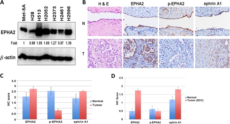
Fig. 4. EPHA2 expression in MPM cell lines and in MPM and SCC tumor tissues.
a Lysates of six MPM cell lines and the Met-5A, a mesothelial control cell line, were immunoblotted with EPHA2 antibody. b Immunohistochemistry representative pictures of 65 MPM tumor and nine normal mesothelium samples were used in MPM TMA. c Protein expression quantity of 65 MPM tumor and nine normal mesothelium samples were used in MPM TMA. d Protein expression quantity of 48 NSCLC SQ and 24 adjacent normal samples were used in SSC TMA. H&E, EPHA2, phospho-(p-)EPHA2, and ephrin A1 were stained and scored. N: normal, T: Tumor

