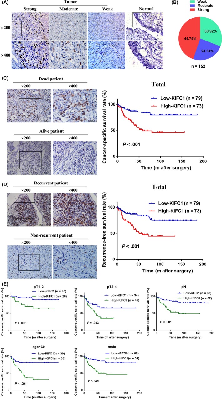
Figure 2.

Upregulation of KIFC1 was closely related to poor prognosis of BC. A, Representative images of KIFC1 staining in paraffin‐embedded BC and normal tissues. Staining intensity in the tumor group was scored as strong, moderate, and weak. KIFC1 was mostly positioned in the nucleus of the bladder specimens. B, Distribution of KIFC1 staining intensity in 152 BC samples. C and D, Kaplan‐Meier cancer‐specific survival (CSS) and recurrence‐free survival (RFS) analysis in the entire cohort of BC patients. Representative immunohistochemical images were also shown. E, Kaplan‐Meier subgroups survival analysis revealed that BC patients with high KIFC1 expression had a lower CSS in different T stages, pN−, age > 60 and male
