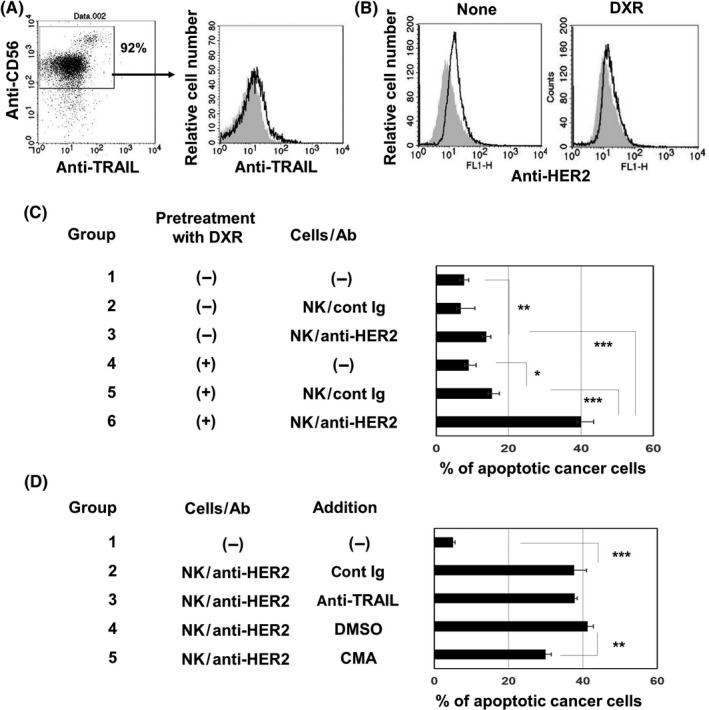
Figure 4.

Perforin is involved in increased sensitivity of doxorubicin (DXR)‐treated MDA‐MB‐231 cells to cytotoxicity by natural killer (NK) cells and anti‐ human epidermal growth factor receptor 2 (HER2) Ab. A, Left panel, NK cells prepared from PBMCs of a healthy donor were stained with allophycocyanin‐conjugated anti‐CD56 Ab and phycoerythrin (PE)‐conjugated anti‐tumor necrosis factor‐related apoptosis‐inducing ligand (TRAIL) Ab. Right panel, histogram of TRAIL expression after gating on CD56+ cells is shown. PE‐conjugated mouse IgG was used as a control (gray background). B, MDA‐MB‐231 cells were stained with anti‐HER2‐FITC. FITC‐conjugated mouse IgG was used as an isotype‐matched control (gray background). C, MDA‐MB‐231 cells were treated with or without DXR (250 nmol/L) for 2 d. After harvesting, cancer cells were cultured with prepared NK cells with the indicated Ab for 6 h. After harvesting these cells, whole cells were first stained with anti‐CD45‐APC, followed by annexin V‐FITC and analyzed by flow cytometry, as Figure 2B. Results of 3 wells are shown. *P < .05, **P < .01, ***P < .005. D, Similarly, DXR‐treated MDA‐MB‐231 cells were cultured with prepared NK cells with the indicated Ab for 6 h. In some groups, either anti‐TRAIL Ab, control mouse IgG, concanamycin A (CMA), or DMSO as a vehicle control was added 1 h before the addition of cancer cells. After harvesting these cells, whole cells were first stained with anti‐CD45‐APC, followed by annexin V‐FITC and analyzed by flow cytometry, as Figure 2B. Results of 3 wells are shown. **P < .01, ***P < .005
