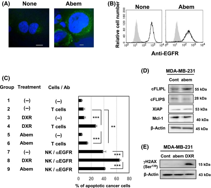
Figure 5.

Abemaciclib treatment does not increase the sensitivity of MDA‐MB‐231 cells to immune cell‐mediated cytotoxicity. A, Confocal imaging was carried out on untreated or abemaciclib‐treated MDA‐MB‐231 cells. Scale bar = 10 μm. B, MDA‐MB‐231 cells were stained with anti‐epidermal growth factor receptor (EGFR) Ab‐FITC. Gray background shows staining with FITC‐conjugated mouse IgG as a control. C, MDA‐MB‐231 cells were treated without or with doxorubicin (DXR; 250 nmol/L) or abemaciclib (Abem; 1 μmol/L) for 48 h. After harvesting, cancer cells were cultured with activated T cells and prepared natural killer (NK) cells and with anti‐EGFR Ab for 6 h. After harvesting these cells, whole cells were first stained with anti‐CD45‐APC, followed by annexin V‐FITC and analyzed by flow cytometry, as Figure 2B. Results of 3 wells are shown. **P < .01, ***P < .005. D, MDA‐MB‐231 cells were cultured with or without abemaciclib (1 μmol/L) for 48 h. Immunoblotting analysis was carried out using the lysates with the indicated Abs. β‐Actin was used as a control. E, MDA‐MB‐231 cells were cultured with or without abemaciclib (1 μmol/L) or DXR (250 nmol/L) for 48 h. Immunoblotting analysis was undertaken using anti‐γH2AX Ab. β‐Actin was used as a control
