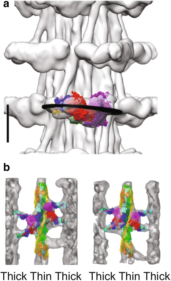
Fig. 5.

The Lethocerus thick filament and troponin bridges. a Cryo-EM image of a relaxed IFM thick filament. Myosin heads form crowns spaced at 14.5 nm along the filament. The IHM is nearly perpendicular to the filament axis, with the blocked head projecting furthest from the filament. The bare zone of the filament is at the top. Scale bar 10.0 nm. b Averaged images of IFM during isometric contraction. Crossbridges are fitted with atomic models of a myosin head and Tn bridges are not. Tn is orange and Tm is yellow. Tn bridges contact the thin filament at or near Tn. a is from Hu et al. (2017); b is from Wu et al. (2010)
