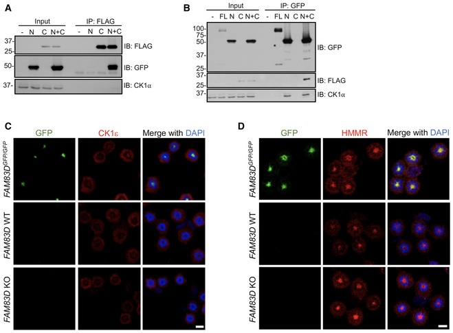
Figure EV2. Exploring the mechanism and specificity of the FAM83D–CK1 interaction.

-
A, BFAM83D −/− U2OS cells were transiently transfected with plasmids encoding GFP‐tagged FAM83D N‐terminus incorporating the DUF1669 (N), FLAG‐tagged FAM83D C‐terminus (C) domain lacking the DUF1669, or with both N and C fragments together; as an additional control, full‐length (FL) FAM83D was also included in panel (B). Cells were lysed and extracts subjected to anti‐FLAG (A) or anti‐GFP (B) immunoprecipitations (IP). Whole‐cell extracts (input) and IP samples were separated by SDS–PAGE, before immunoblotting (IB) with the indicated antibodies.
-
C, DWild‐type (WT), FAM83D −/− knockout (KO) and FAM83D GFP/GFP knockin U2OS cells were synchronised in mitosis with STLC, before being subjected to anti‐CK1ε (C) or anti‐HMMR (D) immunofluorescence and GFP fluorescence microscopy. DNA is stained with DAPI. Scale bars, 20 μm.
