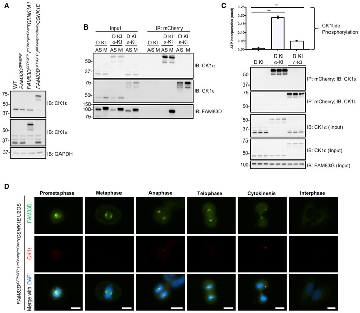
Figure EV4. Verification of mCherry/mCherry CSNK1A1 and mCherry/mCherry CSNK1E U2OS knockin cells.

- Wild‐type (WT), FAM83D GFP/GFP knockin (D‐KI), FAM83D GFP/GFP knockin with mCherry/mCherry CSNK1A1 (D‐KI, α‐KI) and FAM83D GFP/GFP knockin with mCherry/mCherry CSNK1E (D‐KI, ε‐KI) U2OS cells were lysed and subjected to immunoblotting (IB) with the indicated antibodies.
- The cell lines described in (A) were synchronised in mitosis with STLC (M) or left asynchronous (AS). Cells were lysed and subjected to immunoprecipitation (IP) with RFP TRAP beads, before immunoblotting (IB) with the indicated antibodies.
- The cell lines described in (A) were lysed and subjected to IP with RFP TRAP beads, and subjected to an in vitro [γ32P]‐ATP kinase assay using an optimised CK1 substrate peptide (CK1tide) and radiolabeled ATP. n = 3, Error bars, SEM, ***P < 0.0001; ANOVA.
- Asynchronous FAM83D GFP/GFP/mCherry/mCherry CSNK1E knockin U2OS cells were fixed and imaged. Representative images from the indicated cell cycle stages are included. Scale bar, 10 μm.
