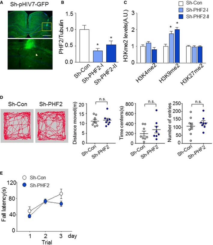
Stereotaxic injection of GFP‐expressing lentiviral particles into bilateral hippocampus CA1 in WT mice. Scale bar, 200 μm.
Quantification of the immunoblot analysis. PHF2 protein levels of hippocampal tissues in sh‐PHF2 (I, II) mice were normalized against tubulin and quantified as fold change relative to that seen in sh‐Con mice.
The H3Kme2 protein levels in sh‐PHF2 (I, II) mice were normalized against H3 and quantified as fold change relative to that seen in sh‐Con mice.
Open field test shows that basal anxiety and locomotor activity in sh‐PHF2 mice were not altered in comparison with sh‐Con mice. Path traces of single‐trial open field tests were recorded for all sh‐Con and sh‐PHF2 mice (left). Distance moved, time in center, and number of entries were scored (right).
Rotarod test showed no differences in motor activity for sh‐Con and sh‐PHF2 mice.
Data information: In (B, C), data are presented as the mean ± SD (
n = 6). *
P < 0.05 (unpaired, two‐sided Student's
t‐test). In (D, E), data are presented as the mean values ± SEM (
n = 8). Data were analyzed using unpaired, two‐sided Student's
t‐test.

