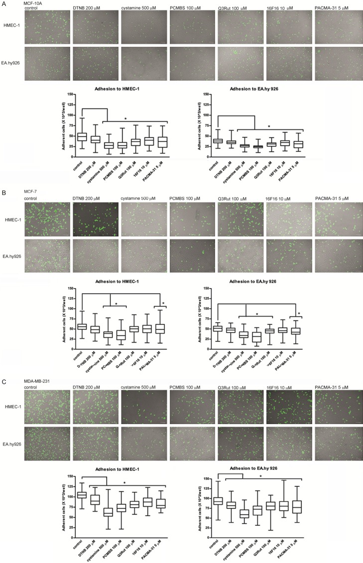
Figure 4.
Adhesion of MCF-10A (A), MCF-7 (B) and MDA-MB-231 (C) Cells to endothelial cells HMEC-1 and EA.hy926 in the presence of PDI inhibitors and free thiol blockers. Data presented as mean + SD; mini-max ranges marked as whiskers; n=10. Total numbers of adherent cells were estimated with a multifunctional plate reader. Significance of differences was analysed with one-way ANOVA and the post-hoc multiple comparisons Tukey’s test and planned comparisons were verified the bootstrap-boosted unpaired student’s t test (10000 iterations) with the Bonferroni’s correction for multiple comparisons.

