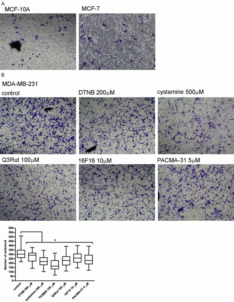
Figure 5.

Migration of MCF-10A, MCF-7 and MDA-MB-231 cells through the gelatin-coated transwell chamber. Data presented as mean + SD; mini-max ranges marked as whiskers; n=5. MCF-10A, MCF-7 (A) and MDA-MB-231 cells (B) Were incubated with PDI inhibitors and free thiol blockers following their prior stimulation with 5 mg/ml LPS and allowed to migrate through the gelatine-coated chamber for 24 hr. Significance of differences was analysed with one-way ANOVA and the post-hoc multiple comparisons Tukey’s test and planned comparisons were verified the bootstrap-boosted unpaired student’s t test (10000 iterations) with the Bonferroni’s correction for multiple comparisons.
