Figure 1.
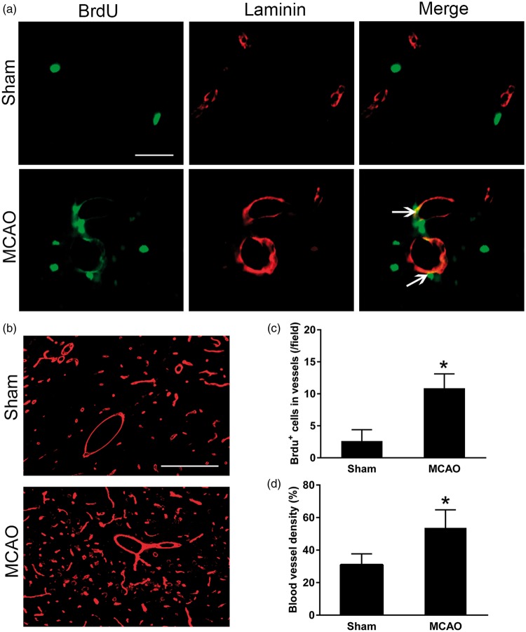
Enhanced angiogenesis in the ipsilateral thalamus after cortical infarction. (a) Immunostaining showing increased expression of BrdU+ cells (green) in laminin-labelled blood vessels (red) in the ipsilateral thalamus at seven days after MCAO. Scale bar: 50 µm. (b) Laminin-immunopositive blood vessels (red) within thalamus in sham-operated controls and ischemic group at seven days after MCAO. Scale bar: 100 µm. (c and d) Quantitative analysis of number of BrdU+ cells that co-stained for laminin and density of blood vessel. n = 8, data are expressed as median ± interquartile range. *P < 0.05 compared with the sham-operated controls.

